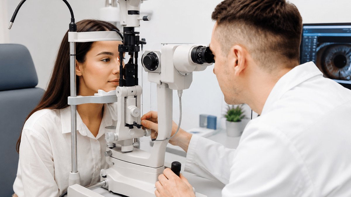
Keratoconus poses a significant challenge to eye care systems in the Middle East, where numerous epidemiological studies have revealed a substantial prevalence of the condition in certain regional populations. A review published in 2022 noted that the highest reported rates of keratoconus have been found in Middle Eastern groups, although the rates vary considerably depending on the population and diagnostic methodology used. Keratoconus is characterized by a progressive thinning and irregularity of the corneal tissue, leading to the development of irregular astigmatism, blurred vision, glare, ghosting, and reduced visual quality in affected patients.
A study conducted among adolescents and young adults in eastern Saudi Arabia found that 2.75% of the participants had keratoconus, while an additional 11.93% were suspected to have the condition. Similarly, a study involving Emirati adolescents estimated the prevalence of keratoconus to be around 2.7%, with a larger group classified as suspected cases. These findings suggest that screening for keratoconus in secondary school populations may be feasible in high-risk settings.
However, it is essential to note that these figures should not be taken as representative of the entire Middle Eastern region, but rather as an indication that keratoconus can affect young people in the region at significant rates, and delayed detection can lead to further progression of the disease before appropriate care is provided.
Early diagnosis is key in managing keratoconus, as it allows for timely intervention and can significantly impact the course of the disease. Modern corneal imaging techniques, such as topography and tomography, have enabled clinicians to detect subtle changes in corneal curvature, elevation, and thickness that may not be apparent through conventional refraction alone. Factors such as family history, persistent eye rubbing, allergies, and unexplained changes in corneal shape can also increase the likelihood of keratoconus and warrant further evaluation. Contemporary reviews describe keratoconus as a multifactorial condition, involving both genetic and environmental influences, rather than a single-cause disease. Early identification provides clinicians with a valuable opportunity to determine the stability and progression of the disease, allowing for more effective management.
Distinction between stabilization and rehabilitation
In the management of keratoconus, it is essential to differentiate between stabilizing the condition and improving vision. This distinction is a critical concept in modern keratoconus care. When disease progression is documented, corneal collagen cross-linking may be considered to increase biomechanical stability and reduce the risk of further ectatic progression. However, stabilizing the cornea does not necessarily restore normal optics, and a patient with a successfully stabilized cornea may still experience significant irregular astigmatism, ghost images, glare, or poor night vision.
Conversely, a specialty contact lens can provide excellent functional vision without altering the biological progression of the disease. Effective keratoconus management requires clinicians and patients to address two distinct questions: What does the patient need to see and function well, and what does the patient need to prevent further disease progression?
Visual rehabilitation is a critical component of keratoconus management, and it involves more than just correcting visual acuity. Glasses and conventional soft contact lenses may provide satisfactory vision in mild cases, but as corneal irregularity increases, conventional refractive correction becomes less effective. Rigid gas-permeable, hybrid, and scleral lenses can help create a more regular optical surface, with scleral lenses being particularly important for patients with moderate or advanced irregular corneas.
These lenses vault over the central cornea and rest on the scleral surface, with a fluid reservoir between the lens and the cornea. A systematic review published in 2025 found consistent improvements in best-corrected visual acuity and vision-related quality-of-life measures among keratoconus patients using scleral lenses, although the authors noted limitations in the available long-term evidence.
Beyond visual acuity: the importance of visual quality
A conventional eye chart only measures a patient’s ability to identify small letters, but it does not provide information on how the patient perceives real-world visual stimuli, such as headlights at night. Patients with irregular corneas may achieve relatively good visual acuity while still experiencing halos, glare, starbursts, reduced contrast, or ghost images. Higher-order aberrations can contribute to these visual disturbances.
Recent research on contact lens correction for keratoconus highlights the role of residual wavefront error and the potential for more sophisticated lens modifications, including wavefront-guided correction, in selected cases. For these patients, the clinical endpoint should not be solely focused on achieving 20/20 vision, but rather on ensuring that the patient’s vision is stable, comfortable, and usable in daily life.
An integrated eye care pathway is essential for effective keratoconus management. This approach involves more than just treating individual eye care interventions in isolation. Keratoconus is a prime example of why integrated eye healthcare services are key. The ophthalmologist may need to evaluate disease progression and determine the need for medical or surgical intervention, while the optometrist specializing in irregular corneas focuses on functional visual rehabilitation and specialty lens fitting. Corneal imaging provides objective structural information, and wavefront evaluation can help explain residual visual-quality complaints. Long-term follow-up connects these pieces over time, ensuring that each stage of the care pathway addresses the correct clinical question.
Regional access to advanced corneal rehabilitation
As healthcare systems in the Gulf and wider Middle East invest in increasingly sophisticated diagnostic and specialty services, patients are becoming more mobile in their search for complex care. M’Eye Clinic in Jerusalem, Israel, is an example of a regional center focused on advanced optometric management of irregular corneas and complex visual rehabilitation.
The clinic works with keratoconus, post-corneal-transplant cases, severe ocular surface conditions, and difficult specialty lens fittings, including customized scleral lens technologies. It also receives international patients from countries such as the UAE, Saudi Arabia, Qatar, Bahrain, and Kuwait. The relevance of centers like this lies not in the fact that every keratoconus patient requires highly advanced technology, but rather in their ability to provide specialized care when standard correction is no longer effective.




Leave a Reply